


What is foot arthritis?
The mid-foot (Talo-Navicular and Calcaneo-Cuboid) joints are responsible for forefoot rotation. The hind-foot (subtalar) joint is responsible for side to side movement of the heel. These joints allow us to walk on uneven ground.
Arthritis is a process of wear and tear involving one or more of these joints. It is commonly a consequence of a previous injury or fracture or a mal- alignment of the foot joints. Patients with rheumatoid disease can also suffer arthritis of these joints. Sometimes it happens without any obvious cause.
Patients usually complaint of pain that becomes worse with walking (particularly on un-even ground), together with stiffness, recurrent swelling and a sense of giving way.
The diagnosis is made by clinical examination and x-rays. A CT-scan is sometimes needed to show the joints involved in even more details.
Mid and Hind-foot Joint Fusions
Why do you need this operation?
Before being offered this operation, you should have tried other measures and treatments as footwear changes, using cushioned inserts, braces and walking aids, taking anti-inflammatory medications and limiting ‘impact’ activities.
This operation is offered to you if you continue to have symptoms despite trying the above measures and after you have been examined and counseled.
The operation is usually offered in severe cases of arthritis of these joints.
What does the operation involve?
The operation is done as an inpatient and you will be discharged when mobile and comfortable.
The operation is performed with general anaesthetic and nerve block (which means numbing the nerves of the foot and ankle).
The operation involves two incisions over the sides of the ankle / hind-foot.
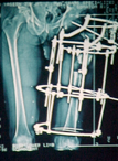
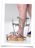
The remaining cartilage covering the joint surfaces is removed and the ends of the joints are fixed using screws, plates, metal staples or metal wires.
Your leg is placed in a plaster.
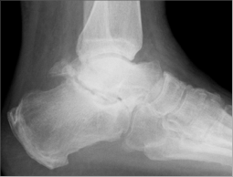
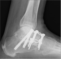
Before After
Your post-operative period
-
•You see the physiotherapist after your operation and they give you crutches
-
•You must keep your foot raised for the first two weeks and when necessary afterwards
-
•Use crutches for the first six weeks.
Your follow-up
-
•Clinic at two weeks to remove the initial plaster and stitches. A full below knee non-weight bearing plaster is then applied
-
•Clinic at six weeks to remove the plaster and take x-rays. A new full below knee weight bearing plaster or removable boot is then applied
-
•Clinic again at 12 weeks to remove the plaster
-
•Off work for around 12 weeks, depending on your job
-
•No driving for 12 - 14 weeks.
What are the possible complications?
-
•Infection
-
•Ongoing pain
-
•Failure of bone healing (non-union)
-
•Sensitive or painful scars
-
•Clots in the leg (DVT)
-
•Clots in the lung (PE)
-
•Chronic Regional Pain Syndrome
Smoking, diabetes, rheumatoid arthritis or being on steroids or blood-thinning medication increases possible risks significantly.
Prophylaxis against clots:
According to the current guidelines you could be prescribed blood thinning medication to reduce your risk of getting clots. You will usually be prescribed two tablets of “Aspirin” to take daily (150 mg). In some cases you will receive “Low Molecular Weight Heparin” subcutaneous (under the skin) injections. You will be shown how to self inject once daily.
Foot Arthritis

Mr. Hisham Shalaby

Consultant Orthopaedic Surgeon
2-8 Millar Crescent Morningside
Edinburgh
EH10 5HW
Telephone: 0131 446 3048
Fax: 0131 447 5778